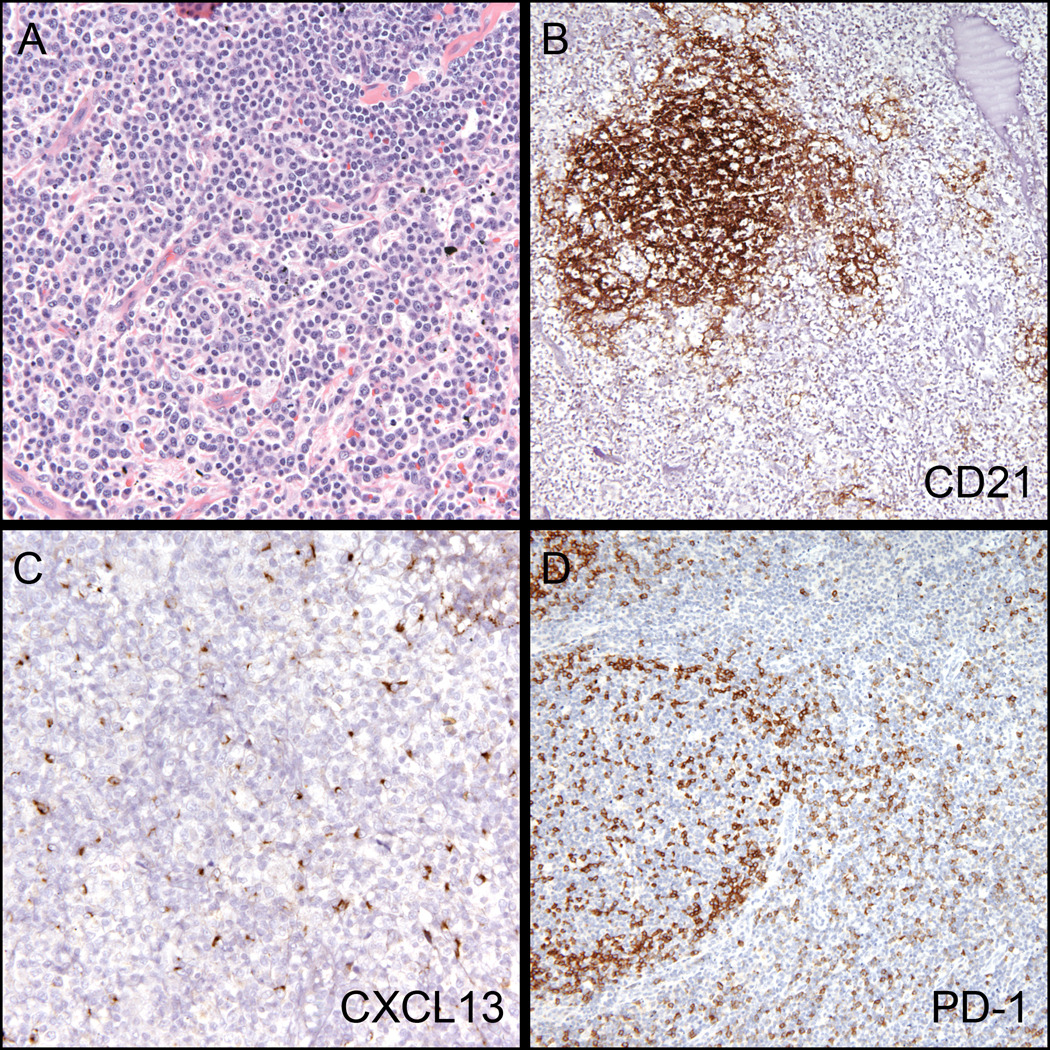
Figure 1. Typical morphologic and immunohistochemical findings in Angioimmunoblastic T-cell lymphoma.

(A) Diffuse paracortical expansion comprised of atypical lymphocytes, eosinophils and prominent blood vessels (H&E, 200x). (B) Expanded follicular dendritic cell network (CD21, 100x). (C) Increased labeling with CXCL13 within the paracortex (CXCL13, 200x). (D) Increased labeling with PD-1 within the paracortex – note normal pattern in germinal center. (PD-1, 200x).
