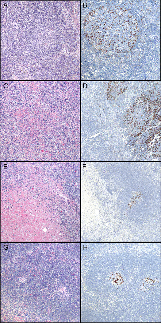
Figure 2. PD-1 staining patterns in non-neoplastic or reactive lymphoid conditions.
(A,B) Reactive follicular hyperplasia (H&E, left, 100x). Note accentuation at the periphery of the germinal center and few positive cells in the mantle zones (PD-1, right, 200x). (C,D) Cat-scratch disease (H&E, left, 100x & PD-1, right, 100x). (E,F) Kikuchi lymphadenitis (H&E, left, 100x). Note absence of staining within regions of necrosis (PD-1, right, 100x). (G,H) Castleman Disease, hyaline vascular type (H&E, left 100x). Note atrophic follicles with dense collection of PD-1 staining (PD-1, right, 100x).

