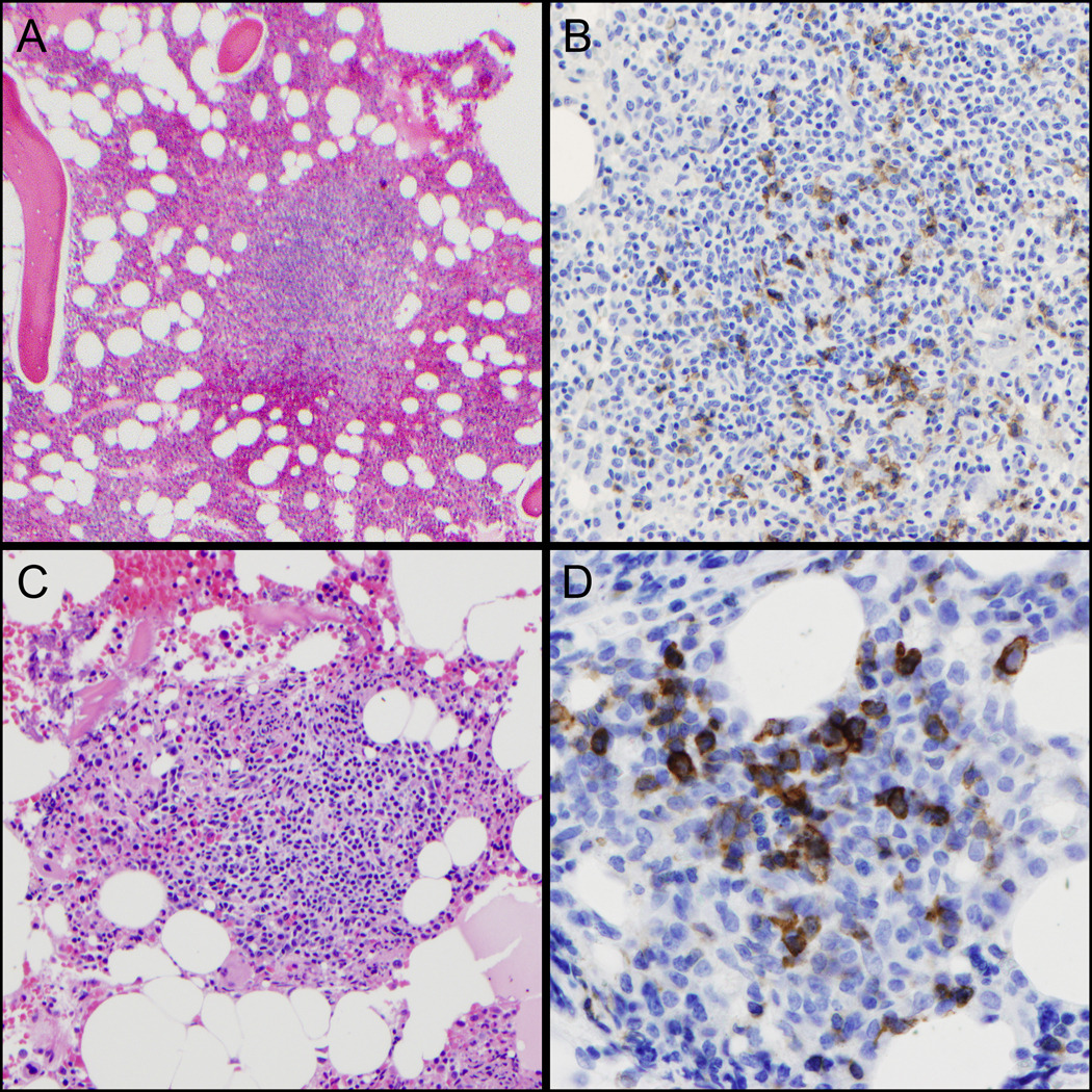
Figure 3. PD-1 staining in bone marrow biopsies.

(A,B) Reactive lymphoid aggregate with germinal center. PD-1 (right) stains scattered small GC-T-cells in the area of the germinal center (H&E, left, 100x & PD-1, right, 200x). (C,D) Bone marrow involvement by AITL. The lymphoid aggregate contains a mixture of eosinophils and branching vessels (H&E, left, 200x). PD-1 (right) is strongly positive in the large, atypical lymphoma cells (right, 400x).
