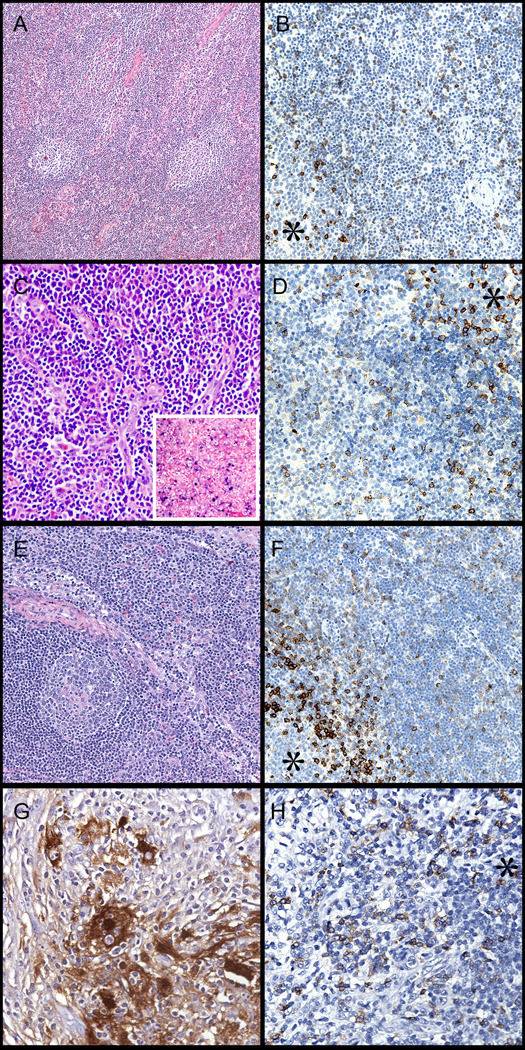
Figure 5. Reactive conditions showing an abnormal pattern of PD-1 expression.
* Asterisk designates a germinal center. (A,B) Atypical paracortical hyperplasia in 22 year old female. Note the presence of reactive germinal centers and increased vasculature in the paracortex (H&E, left, 100x). PD-1 labels many cells outside of follicles. (PD-1, right, 200x). (C,D) Infectious mononucleosis in 19 year old male (H&E, left, 200x & EBV in-situ hybridization, inset). PD-1 labeling in the paracortex, with germinal center (asterisk) for comparison (right, 200x). (E,F) Lymph node in HIV infected patient (H&E, left, 100x). PD-1 labeling in paracortex with germinal center for comparison (right, 200x). (G,H) Rosai-Dorfman Disease in the soft tissue of a 41-year old man (S100 stain, left, 400x). Note the typical histiocyte of this disorder and the presence of emperipolesis. PD-1 stains many cells throughout the lesion (right, 200x).

