
FIG. 2.
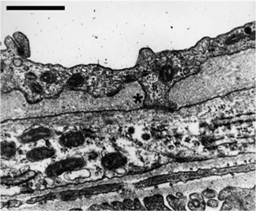
Myoendothelial junction in mouse cremaster arteriole. Usually, the use of electron microscopy is required to identify the MEJ in arterioles. In this electron micrograph, an EC extension is seen breaking through the internal elastic lamina and making contact with an SMC at the MEJ (*). Bar is 1 μm.