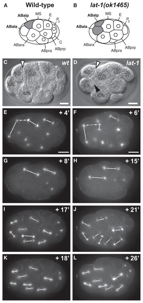
Figure 3. Division Plane Defects of lat-1 Mutants.
Left column, WT embryo; right column, lat-1 embryo. (C and D) DIC microscopy; (E–L) spindle orientation visualized by GFP::β-tubulin. Centromere pairs are linked with white lines. Time points are shown in minutes relative to AB4 stage (Figures S3C and S3D). (A–D) Relative positions of ABala, ABalp, and MS in WT (A and C) and lat-1 (B and D) embryos. Note that in lat-1 ABala forms an extensive membrane interface with MS (black arrowhead in [D]). (E) a-p alignment of ABal and MS spindles in WT embryo. (F) In lat-1 embryo a-p spindle alignment fails in ABal and is delayed in MS. a-p alignment of E precedes MS. The distance between the posterior ABal centromere and anterior MS centromere is marked by double arrows. (G and H) Delayed a-p alignment of MS spindle in mutant (H) compared to WT (G). (I–L) Rapid a-p alignment of AB8 spindles in WT embryos (I and K). Delayed alignment in lat-1 embryos (J and L) and perpendicular division planes of sister cells. In all embryos anterior is to the left, posterior is to the right, and ventral (MS) is up. Scale bars = 5 μm.

