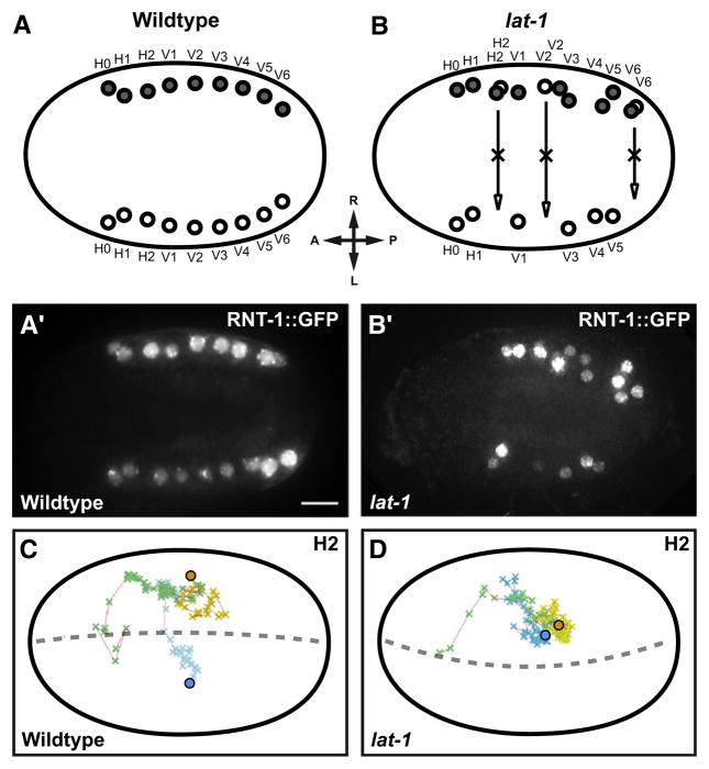
Figure 4. Seam Cell Positioning Defects in lat-1 Mutants.
(A and A′) The seam cell marker RNT-1::GFP labels two seam cell rows of ten cells each on either side of WT embryos.
(B and B′) RNT-1-positive cells persist on the right side in lat-1 mutant.
(C and D) The migration path of H2 seam cells and their precursors was traced by 4D microscopy. Green, migration path of common ABarpp precursors. Movement of ABarpp descendants after left-right split of cell lineage: blue, left descendants (ABarppax); yellow, right descendants (ABarpppx); spheres, final destination of differentiated seam cells. (C) Contralateral migration of H2L/H2R in WT embryo. (D) In a lat-1 embryo left-right migration is absent, and H2L and H2R stay together. Dorsal views, anterior to the left. Scale bar = 5 μm.

