
Fig. 4. CTIP1 potentiates the ARP1-mediated repression in a TSA-insensitive manner.
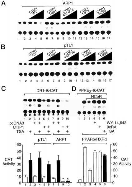
A and B, HEK 293 cells were cotransfected with 5 μg of the DR1-TK-CAT reporter, 5 μg of Myc-ARP1 expression vector (A) or empty vector (pTL1; B) and increasing amounts (0.2, 0.66, and 2.22 μg) of expression vectors encoding full-length HA-CTIP1 or HA-CTIP1 deletion mutants as indicated. CAT reporter activity in cell extracts was determined as described previously (21). C, HEK 293 cells were cotransfected with 5 μg of DR1-TK-CAT reporter, 5 μg of Myc-ARP1 expression vector or empty vector (pTL1), and 2.2 μg of HA-CTIP1 or empty vector (pcDNA3). Cells were treated with TSA (100 ng/ml; solid bars) as indicated for 24 h prior to harvesting and CAT assays. D, cotransfection of HEK 293 cells with 2 μg of PPRE-TK-CAT reporter and expression vectors for PPARα/RXRα (0.5 μg each) and NCoR (2 μg) as indicated. Cells were treated with either vehicle (0.1% Me2SO; lane 3); the RXR agonist, 9-cis-retinoic acid (9cRA; 1 μm); or the PPARα agonist, WY-14,643 (10 μm), or TSA as noted for 24 h prior to harvesting and determination of CAT activity. The quantifications shown below B and C represent mean CAT activities ± S.D. derived from three independent experiments. The CAT activity values in lanes 9 and 10 are statistically different from those shown in lanes 7 and 8, respectively, as determined by Student’s t test (p < 0.05, indicated by asterisks).