
Fig. 5. Localization of HA-CTIP1 (A) and Myc-ARP1 (C) in HEK293 cell nuclei.
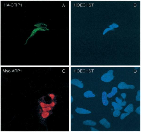
HEK293 cells growing on coverslips were transiently transfected with expression vectors encoding HA-CTIP1 (A) and Myc-ARP1 (C). Forty-eight hours after transfection, the cells were fixed and incubated with anti-HA (A) or anti-Myc (C) antibodies and stained with appropriate fluorescein isothiocyanate- or tetramethylrhodamine isothiocyanate-conjugated secondary antibodies detecting HA-CTIP1 and Myc-ARP1 immune complexes, respectively. B and D, Hoechst-counterstained cells shown in A and C, respectively. HA-CTIP1 exhibited the punctate staining pattern depicted in A in 80% of transfected cells examined, with the balance displaying a combination of focal and diffuse staining (see Table I). In contrast, Myc-ARP1 exhibited the diffuse staining pattern in 100% of 120 transfected cells examined by a naive observer. Images were obtained on a Leica inverted confocal microscope model TCS4D using a × 100 objective. The images shown are derived from a representative experiment that was replicated several times.