
Fig. 6. ARP1 is redistributed in the nucleus when cotransfected with CTIP1.
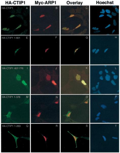
HEK293 cells were transiently cotransfected with expression vectors encoding Myc-ARP1 and either HA-CTIP1 or HA-CTIP1 mutants as indicated. The corresponding proteins were localized 48 h after transfection by indirect immunofluorescence confocal microscopy. Cells were stained for HA-CTIP1 (first column) and Myc-ARP1 (second column) as described in the legend of Fig. 5. An overlay of the images presented in the first two columns is shown in the third column, and the fourth column represents counterstaining of the cells with Hoechst as indicated. Shown are representative experiments that were replicated 3–7 times. Each micrograph was prepared using a × 100 objective on a Leica inverted confocal microscope model TCS4D, and overlays were prepared using Photoshop 5.0.