
Figure 7.
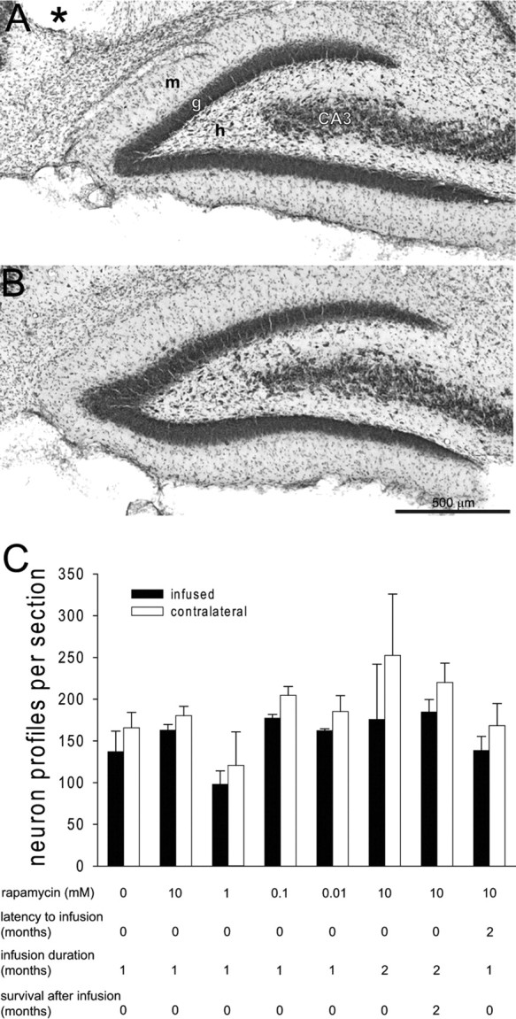
Rapamycin infusion did not protect hilar neurons from status epilepticus-induced loss. Nissl stained dentate gyrus after 1 month infusion with 10 mm rapamycin (A) and contralateral noninfused hippocampus (B). The asterisk indicates the infusion site. m, Molecular layer; g, granule cell layer; h, hilus; CA3, CA3 pyramidal cell layer. C, The average number of hilar neuron profiles per section in the three sections closest to the infusion site and corresponding sections of contralateral hippocampus for all experimental groups. The number of hilar neuron profiles was slightly and consistently lower in infused than in contralateral hippocampi (p = 0.006, ANOVA), regardless of experimental group. Error bars indicate SEM.