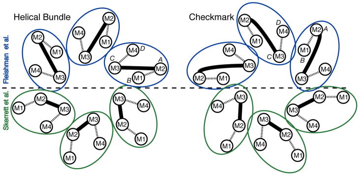
Figure 2.
Possible molecular boundaries for the connexin subunit include a helical bundle (left) and a “checkmark” (right) (following the naming of Unger et al., 1999). Each shows two assignments for the 4 TM α-helices within each subunit, according to Fleishman et al. (2004) (top, blue) and Skerrett et al. (2002) (bottom, green). Dashed lines denote the extracellular loops, E1 and E2, and the solid lines denote the M2–M3 cytoplasmic loops. The α-helical rods designated A, B, C and D in the 3D density map derived by electron cryo-crystallography (Unger et al., 1999) are also indicated.

