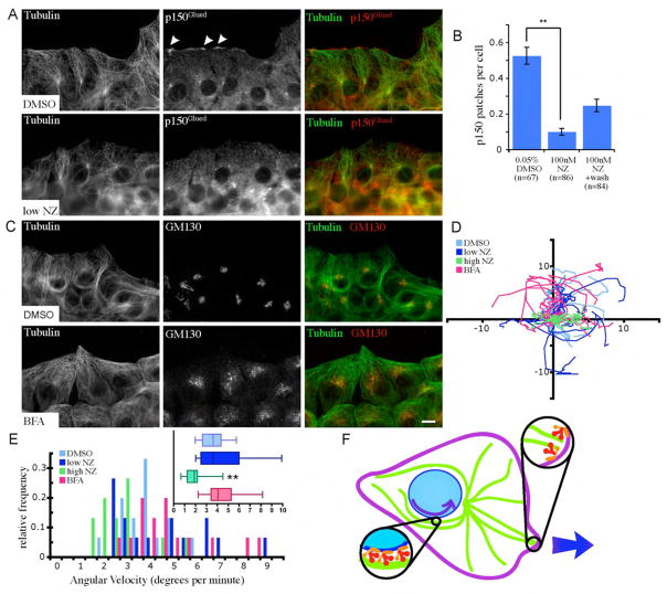
Figure 6.
Nuclear rotations are independent of leading-edge- and Golgi-localized dynein. (A) Cells treated with low doses of nocodazole (NZ) 5.5 hours after wounding maintain the MT network morphology (green), but lose patches of p150Glued (red) at the leading edge. (B) Dynactin patch accumulation is inhibited in cells treated with low-doses of NZ. Patches are partially rescued 30 min after NZ washout. (C) Cells treated with BFA 5.5 hours after wounding maintain MT network morphology (green) but have fragmented Golgi (red, Golgi marker GM130). Bar, 10 microns. (D) Tracks of nucleoli treated with DMSO, BFA, or NZ during migration of fibroblasts over 45 min. Axis labels are in microns. (E) Angular velocity nuclei of cells treated with DMSO (control), NZ, or BFA (n=15). (F) Scheme for two roles for dynein/dynactin in cell motility: (1) Dynein and dynactin accumulate in cortical patches at the leading edge, where they interact with MTs and mediate centrosome and MT orientation during motility, and (2) dynein and dynactin interact with the nuclear envelope and transport the nucleus along MTs on the sides of the nucleus. Error bars indicate SEM; ** indicates p<0.005.

