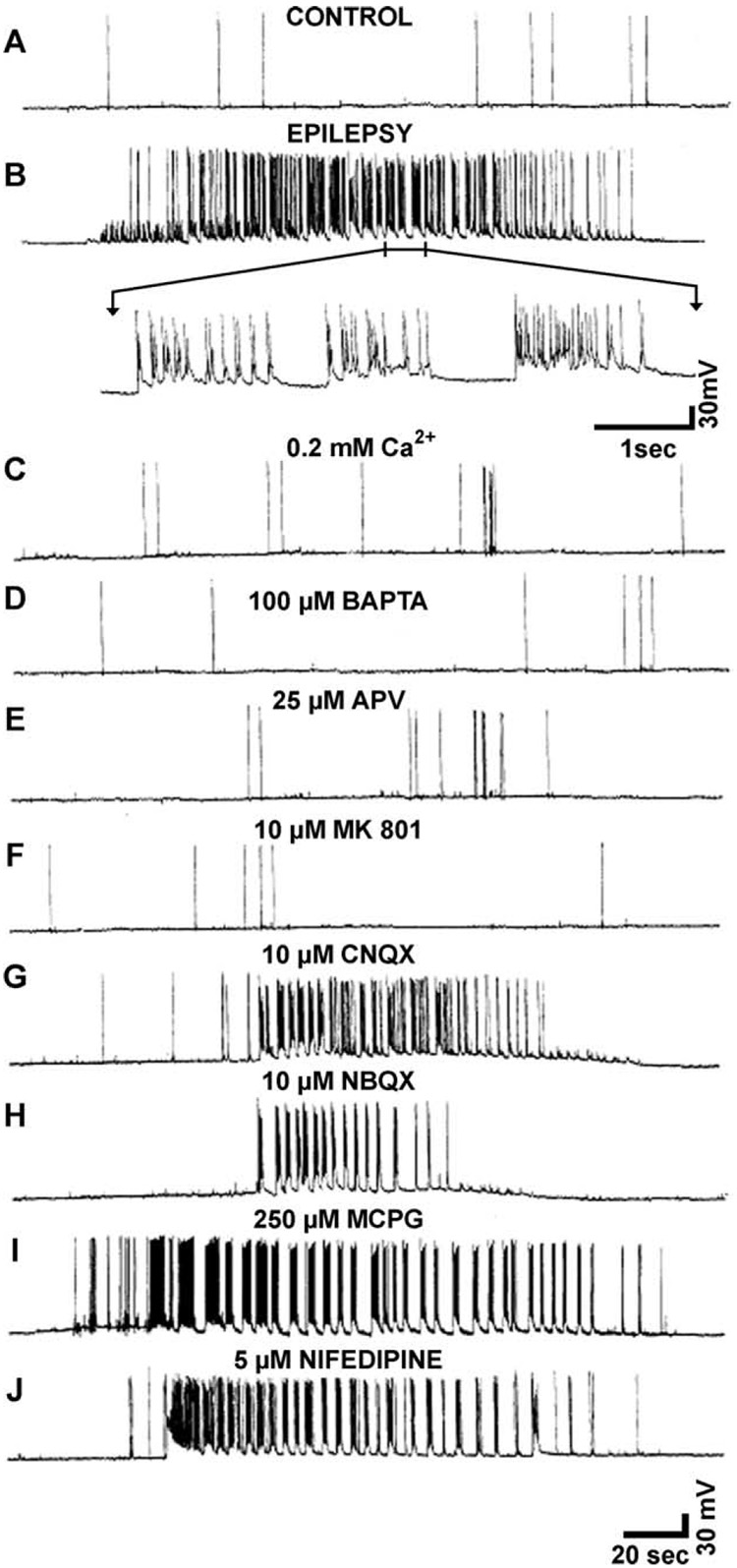
Fig. 7.
Induction of spontaneous recurrent seizures in cultured hippocampal neurons 2 days after low-Mg2+ treatment in the presence or the absence of low Ca2+ (C) or neuropharmacological agents (D–J) during the brief 3-hr exposure to low Mg2+. After low-Mg2+ treatment, cultures immediately were returned to normal Mg2+ levels in the media. Intracellular recordings were obtained from hippocampal pyramidal neurons 2 days after low-Mg2+ treatment. The recordings shown were representative of more than 10 independent experiments for each condition. SREDs (seizures) were observed under standard conditions, 2 mM Ca2+ (B), 10 µM CNQX (G), 10 µM NBQX (H), 250 µM MCPG (I), and 5 µM nifedipine (J). Low extracellular Ca2+ (0.2 mM; C) or intracellular chelation of Ca2+ with 100 µM BAPTA (D) during low-Mg2+ treatment completely blocked the development of spontaneous recurrent seizures despite causing even a higher-frequency seizure discharge during the low-Mg2+ treatment. Expansion of a 5-sec interval of seizure activity in panel B demonstrated the numerous spikes associated with each epileptiform burst. More than 80 spike discharges occurred during this 5-sec segment, giving an average spike frequency discharge of 16 Hz during this segment of the seizure shown in (B). The electrophysiological patterns for conditions A–J were representative of recordings taken earlier or later than the 2-day post-SE sample time. Conditions that blocked the induction of spontaneous recurrent seizures never manifested seizure discharges. (From DeLorenzo et al., 1998.)

