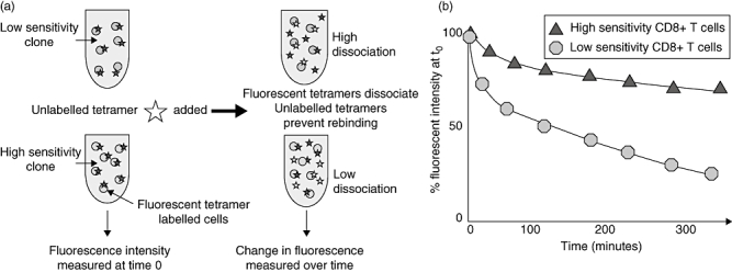
Fig. 2.

Tetramer dissociation assay. (a) Antigen-specific CD8 T cells are stained with fluorescently labelled major histocompatibility complex class I tetramers. Fluorescent intensity is measured at time 0. Unlabelled multimer is added to the tetramer-stained cells after time 0, which prevent rebinding of dissociated labelled tetramers. (b) Change in fluorescent intensity is measured over time. High-sensitivity CD8 T cells demonstrate slower dissociation than low-sensitivity CD8 T cells.
