
Fig. 6.
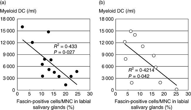
The number of peripheral blood dendritic cells (PBDCs) and tissue dendritic cells (DCs) is related negatively in both primary and secondary SS during clinical time–course. We analysed the number of blood myeloid DCs and examined immunohistochemical staining of biopsied specimens from the 11 patients with primary SS (a) and 10 patients with secondary SS (b). We counted both fascin-positive cells and total MNCs per 100 ×100 µm at least five fields of view (magnification × 100) per each sample. The x-axis indicates mean percentage of fascin-positive cells to total MNCs in labial salivary glands and the y-axis indicates the number of myeloid DCs in blood.