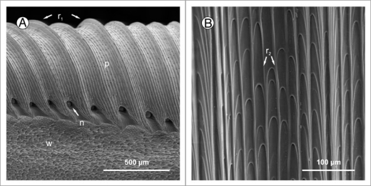
Figure 2.
(A) Peristome surface (p) of Nepenthes alata, structured by first (r1) and second order radial ridges. In between the tooth-like projections at the inner edge of the peristome the pores of large extrafloral nectaries (n) can be seen. Below the peristome is the wax-covered inner wall surface (w). (B) The second order ridges (r2) are formed by straight rows of overlapping epidermal cells.

