
Figure 2.
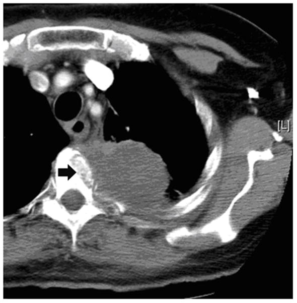
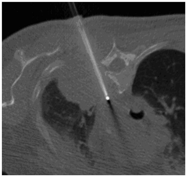
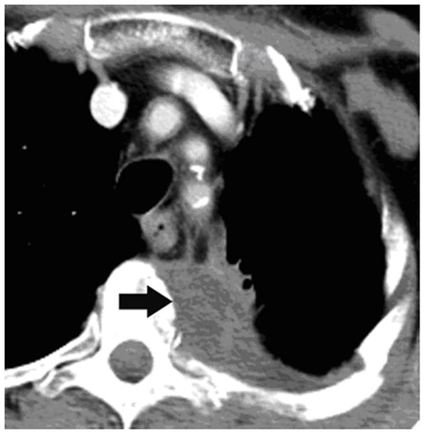
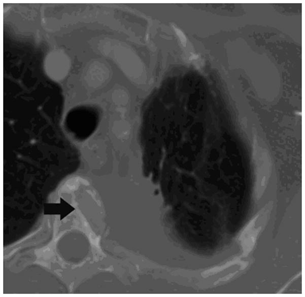
62-year old woman with T4 NSCLC status post prior radiation and chemotherapy. The patient had persistent unremitting pain. Supine CT image (a) shows the large lung mass involving the T4 vertebral body (arrow). RFA of bone-tumor interface was performed under CT-guided fluoroscopy (b,). The patient tolerated the procedure well and her pain improved dramatically. Follow-up CT images 2 years later in soft tissue (c) and bone windows (d) show mass necrosis (arrow) and partial remineralization of the bone destruction (arrow). The patient remains pain free over 2 years after RFA.