
Fig. 1.
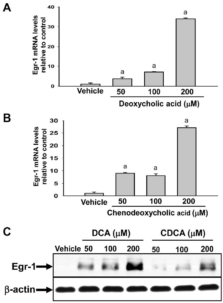
Upregulation of Egr-1 mRNA and protein in primary mouse hepatocytes treated with bile acids. Hepatocytes were isolated from mice and treated with either DCA or CDCA. (A and B) Two hours later, Egr-1 mRNA levels were quantified by real-time PCR. Data are expressed as mean +/- SEM; n=3. aSignificantly different (p < 0.05) from vehicle-treated hepatocytes. (C) Egr-1 and β-actin proteins were detected by western blot. Representative of an n=3.