
Figure 8.
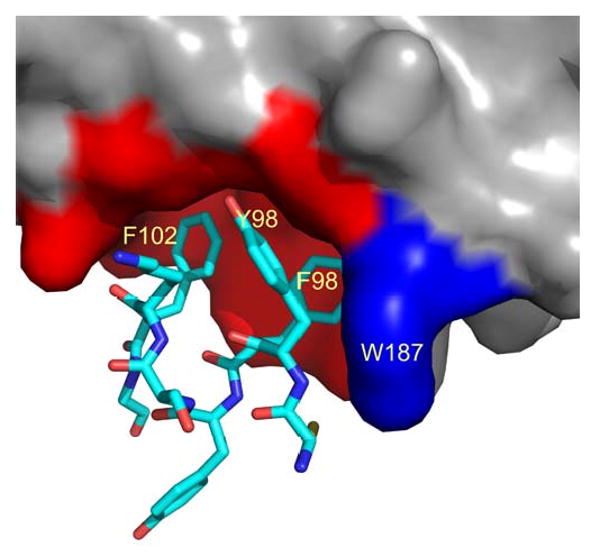
Structure of the interface between NS1-ED and F2F3 of CPSF30. The PDB code is 2RHK. The surface of ED is shown in grey but the receptor pocket (I117, I119,Q121, V180, K108, K110, G183, and G184) is colored red. W187 is shown in blue. Residues 96-103 from F2F3 are shown as sticks.