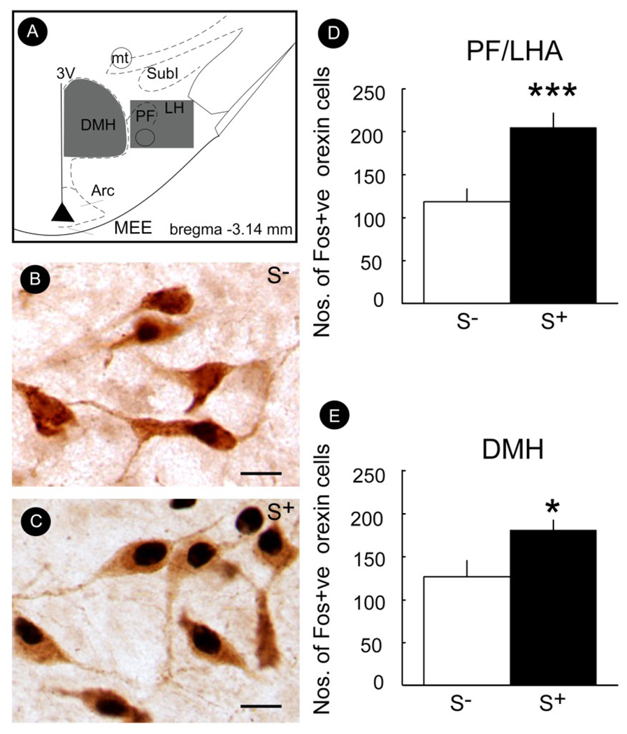
Figure 6.
Mean (± SEM) number of Fos-positive Orx/Hcrt cells in the perifornical/lateral hypothalamus (PF/LHA) (D) or dorsomedial hypothalamus (DMH) (E) in rats exposed to either the ethanol- (S+) or non-reward-related stimulus (S−). A significantly greater number of Fos-positive Orx/Hcrt cells was observed in animals exposed to the S+ (vs. S−) in both the PF/LHA and DMH. (B & C) Photomicrographs illustrating the effects of S− or S+ exposure on the number of Fos-positive Orx/Hcrt cells in the PF/LHA. (A) Schematic illustrating the rostro-caudal level from which photomicrographs (B & C) of PF/LHA tissue, immunolabeled for Fos-protein and Orx/Hcrt, were made. *p < 0.05, ***p < 0.001, compared with S−. Scale bar = 20 µm. Taken with permission from Dayas et al. (2008).

