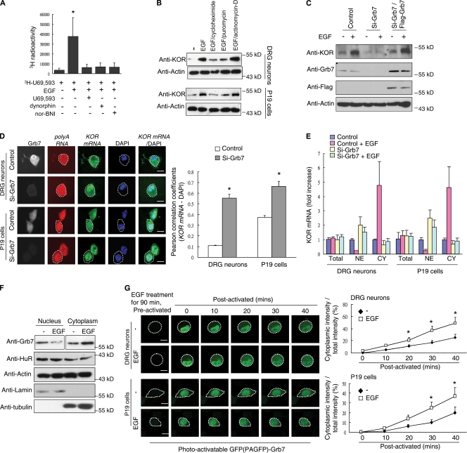
Figure 1.
Grb7 mediates EGF-induced KOR levels and KOR mRNA nuclear export. (A) Quantitative KOR ligand binding assay of primary rat DRG neurons with 3H-U69,593 in the presence or absence of EGF. Cold KOR agonists dynorphin and U69,593 and the antagonist nor-BNI were used for competition (*, P < 0.05). (B) Western blot analysis of primary rat DRG neurons (top) or P19 cells (bottom) in the presence or absence of EGF. Cycloheximide, puromycin, or actinomycin-D was applied along with EGF. (C) Western blot analysis of P19 cells transfected with control siRNA, Grb7 siRNA, or Grb7 siRNA plus an siRNA-insensitive Flag-Grb7 in the presence or absence of EGF. (D) FISH of DRG neurons (top) and P19 cells (bottom) with a fluorescein-12–labeled probe specific to KOR mRNA. Cells were pretransfected with control (first and third rows) or Grb7 siRNA (second and fourth rows). Oligo (dT) was used as a control to hybridize total poly(A) RNA. Silencing of Grb7 was monitored by immunohistochemistry using an anti-Grb7 antibody. Colocalization of KOR mRNA and DAPI was quantified using the Pearson correlation coefficients and is shown on the right (*, P < 0.05). (E) RT-qPCR of KOR mRNA from nuclear or cytoplasmic fractions of rat DRG neurons or P19 cells. Control siRNA– or Grb7 siRNA–transfected cells in the presence or absence of EGF treatment are shown. CY, cytoplasmic fraction; NE, nuclear fraction. (F) Western blot analysis of nuclear and cytoplasmic fractions from P19 cells in the presence or absence of EGF. α-Tubulin and lamin B served as the fractionation controls. (G) PAGFP-fused Grb7 was transfected into DRG neurons (top) and P19 cells (bottom). The images were obtained with a confocal microscope at the indicated time points. The quantified, relative cytoplasmic PAGFP-Grb7 signal from 50 cells is shown on the right (*, P < 0.05). (D and G) White dotted lines outline the cytoplasm of individual cells. (A, D, E, and G) Error bars represent SDs. Bars, 25 µm.

