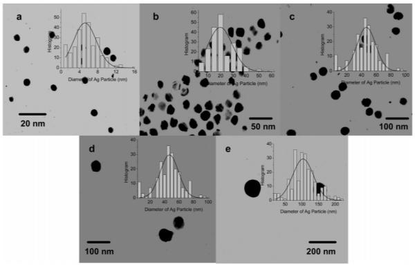
Figure 2.

Transmission electron micrograph (TEM) images of silver particles with the different metal core sizes of (a) 5, (b) 20, (c) 50, (d) 70, and (e) 100 nm. Histograms of the size distributions of the metal particles were inserted correspondingly.
