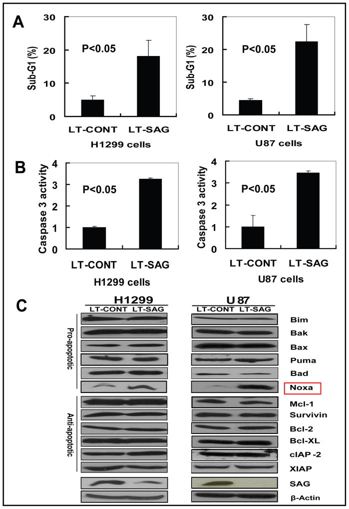
Figure 4. SAG silencing induces apoptosis with Noxa accumulation.
H1299 and U87 cells were infected with LT-SAG, along with LT-CONT for 96 hours, then split and cultured for 72 hours, followed by PI staining and FACS analysis for apoptosis detection, caspase-3 activity assay, cell cycle profile and Western blotting for the levels of apoptosis-associated proteins. (A) Induction of apoptosis by SAG silencing. Apoptotic cells were determined by sub-G1 fraction in FACS analysis; (B) Caspase-3 activation upon SAG silencing. Caspase-3 activity in infected cells was determined by Caspase-3 activity assay. For A and B, shown is value ±SEM of three independent experiments. (C) The expression of apoptosis-associated proteins. The status of a panel of apoptosis-associated proteins including pro-apoptosis proteins and anti-apoptosis proteins were detected by Western blotting with β-Actin as the loading control.

