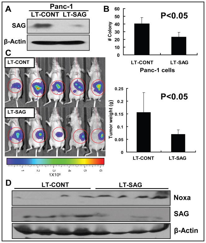
Figure 6. SAG silencing inhibits the growth of orthotopic pancreatic tumors.
PANC-1 human pancreatic carcinoma cells were infected with LT-CONT and LT-SAG, followed by the determination of SAG silencing effect (A), cell survival assay in vitro (B), tumor formation in vivo (C), and Western blotting (D). (A) SAG silencing effects. Panc-1 cells stably transfected with luciferase were infected with LT-CONT or LT-SAG, and the SAG levels were determined 96 hrs post infection by Western blotting with β-actin as the loading control. (B) Clonogenic cell survival assay. Cells after SAG silencing were split, seeded into 6-well plates with 100 cells per well in triplicates, and incubated at 37 °C for 9 days, followed by 0.05% methylene blue staining and colony counting. (C) Bioluminescence imaging of implanted tumors and measurement of tumor weight. Panc-1 cells stably transfected with luciferase were infected with LT-CONT or LT-SAG and implanted into pancreases of the mice (5 mice per group) for the evaluation of tumor growth in vivo. After 5 weeks, tumors were bioluminescence imaged using a cryogenically cooled imaging system coupled to a data acquisition computer running Living Image software at the University of Michigan Small Animal Imaging Core. Mice were then sacrificed, tumors harvested and weighed. (D) NOXA expression in tumors. The level of Noxa, SAG in tumor tissues was determined by Western blotting using antibodies against Noxa and SAG with β-Actin as the loading control.

