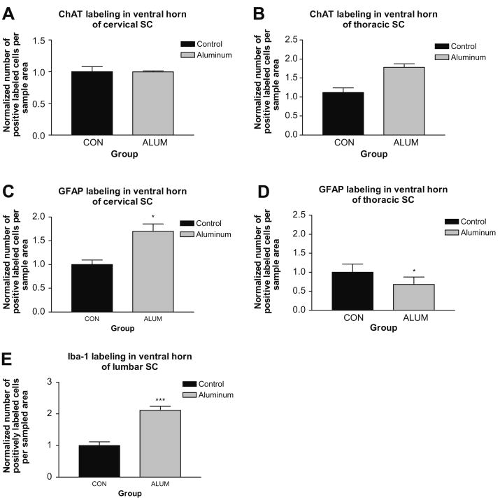
Fig. 1.
Impact of aluminum hydroxide on different levels of spinal cord (SC). (A and B) ChAT labeling in cervical and thoracic cords, respectively. (C and D) Normalized cell counts for GFAP labeling of reactive astrocytes in cervical and thoracic spinal cord, respectively. In cervical cord, the aluminum hydroxide treated groups showed higher levels of GFAP labeling with the aluminum alone group achieving statistical significance. (E) Iba-1 fluorescent labeling in the ventral horn of mouse lumbar cord showed that aluminum-injected mice had significantly increased numbers of activated microglia. Data are means ± S.E.M. ***p < 0.001, one-way ANOVA.

