
Figure 6.
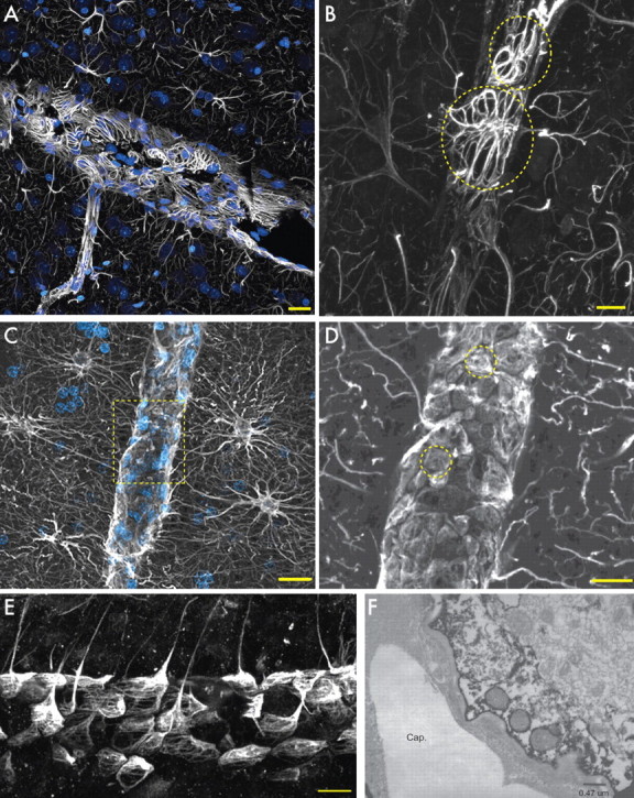
Astrocytic end feet in rodents and humans. A, Cortical blood vessel in rat. GFAP, White; nuclei (DAPI), blue. Scale bar, 20 μm. B, GFAP in end feet forms rosettes on the vessel in the rat. Scale bar, 10 μm. Yellow circles indicate individual end feet. C, Human protoplasmic astrocytes extend processes to the vasculature. Scale bar, 20 μm. D, Yellow box seen in C. GFAP in end feet completely covers the vasculature. Yellow circles indicate individual end feet. Scale bar, 10 μm. E, Transverse section of blood vessel and human astrocyte end feet. Scale bar, 20 μm. F, Electron micrograph of aquaporin 4 immunohistochemistry of the astrocytic end foot on a capillary (Cap.). Note the presence of mitochondria in the end foot of the astrocyte.