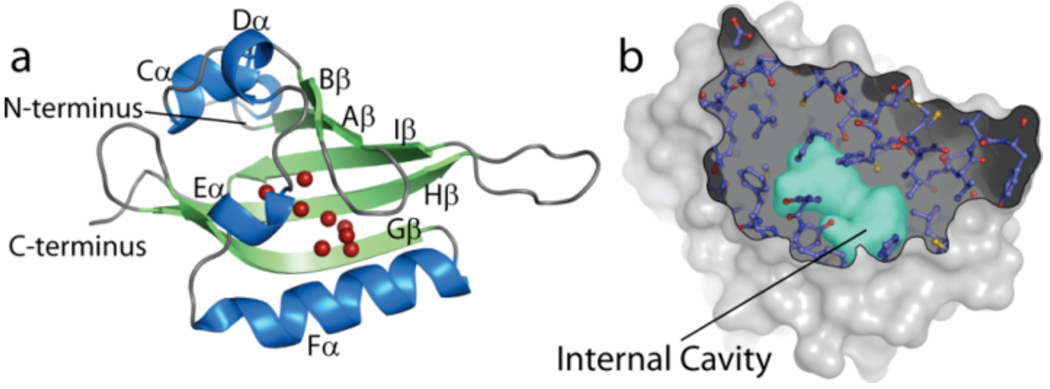
Figure 1. Crystal structures of HIF2α PAS-B reveal and internal solvent-filled cavity which binds artificial ligands.
(a). Crystal structure of the PAS B domain of HIF2α in the apo form showing the eight bound solvent atoms within the core of the domain (red spheres). (b). Cutaway of the HIF-2α surface revealing the 290 Å3 internal cavity, depicted as a surface (cyan).

