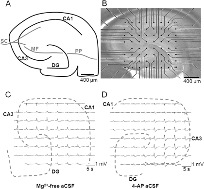
Fig. 1.
Hippocampal slices are amenable to MEA recording. A, schematic representation of hippocampal slice showing the position of CA1, CA3, and DG regions, together with major pathways: Schaffer collateral (SC), mossy fiber (MF), and perforant pathway (PP). B, micrograph showing a hippocampal brain slice (stained with pontamine blue) mounted onto a substrate-integrated MEA (60 electrodes of 30 μm diameter, spaced 200 μm apart in an ∼8 × 8 arrangement). Scale bar, 400 μm. Representative LFP burst activity was recorded at 60 electrodes across a hippocampal slice in (C) Mg2+-free aCSF and (D) 4-AP aCSF. Traces were high pass-filtered in an MC_rack at 2 Hz.

