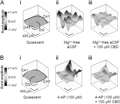
Fig. 3.
CBD attenuates epileptiform activity induced by Mg2+-free and 4-AP aCSF. Representative contour plots illustrating CBD effects upon spatiotemporal epileptiform burst features. A, in the continued presence of Mg2+-free aCSF: quiescent period between epileptiform burst events also showing hippocampal slice orientation (i), peak source in the absence of CBD (ii), and peak source in the presence of CBD (100 μM) (iii). B, in the continued presence of 100 μM 4-AP: quiescent period between epileptiform burst events also showing hippocampal slice orientation (i), peak source in the absence of CBD (ii), and peak source in the presence of CBD (100 μM) (iii).

