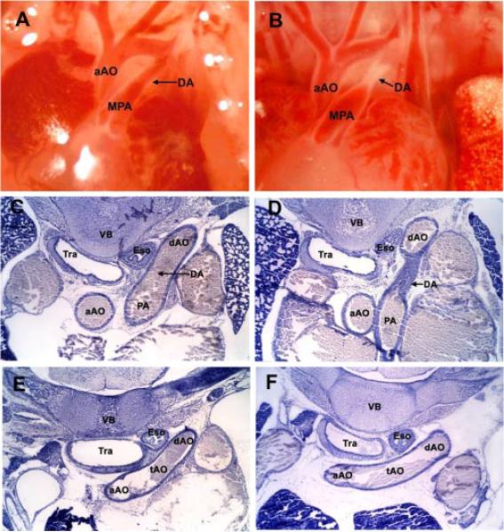
Fig. 2.
Physiological closure of the mouse ductus arteriosus. Anatomical views of the open fetal ductus arteriosus on the morning of day 19 of gestation (A) and constricted newborn ductus arteriosus at 4 h of age (B). Serial thoracic sections demonstrate histologic features and dimensions of the open fetal ductus (C) and adjacent transverse aortic arch (E) compared with the closed newborn ductus (D) and corresponding transverse aortic arch (F). aAO, ascending aorta; tAO, transverse aorta; dAO, descending aorta; PA, pulmonary artery; DA, ductus arteriosus; Tra, trachea; Eso, esophagus, VB, vertebral body.

