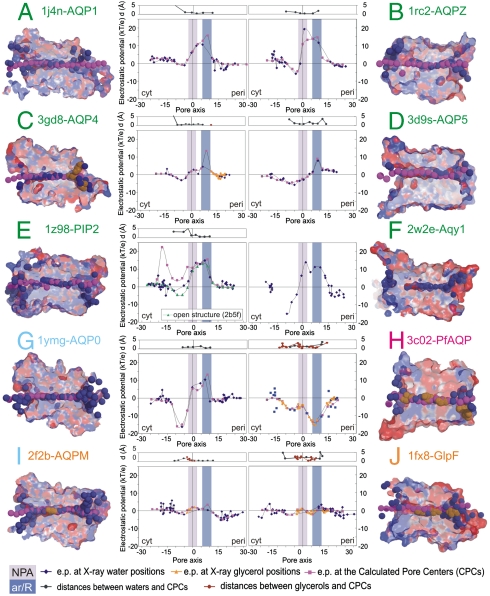
Fig. 1.
Electrostatic potentials into the channels, calculated at the crystallographic positions of water/glycerol molecules and at the pore centers predicted by the PoreWalker program (33). Orthodox aquaporins are labeled in green, the low water conductance AQP0 in cyan, aquaglyceroporins in orange, and the bifunctional PfAQP in magenta. Sections of the corresponding PDB structures are shown colored according to the electrostatic potentials. X-ray water and glycerol atoms inside the channels are shown as deep blue and orange surfaces, respectively. Dummy atoms at the geometrical centers of the pores are shown as magenta surfaces.

