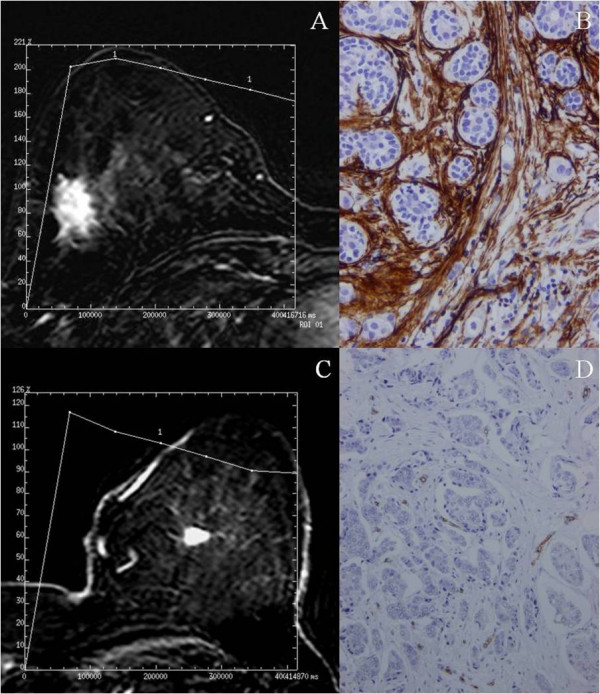
Figure 1.

Breast MR image acquired in a 50-years-old patient with a palpable mass in the right inferior outer quadrant. The axial postcontrast subtracted image (9.9/4.2; flip angle, 10°) depicts a lesion with spiculated margin of mass (arrow). The time-signal intensity curve of this shows a type III time course with a peak of maximal enhancement after two min. (A). Immunohistochemical staining of CD34 in the same tumor showing a high microvessel density. 100×. (B). Breast MR image acquired in a 63-years-old patient with a palpable mass in the left upper inner quadrant. The axial postcontrast subtracted image (9.9/4.2; flip angle, 10°) depicts a lesion with smooth margin of mass (arrow). The time-signal intensity curve of this shows a type III time course with a peak of maximal enhance before 2 min (C). Immunohistochemical staining of CD34 in the same tumor showing a low microvessel density. 100×. (D).
