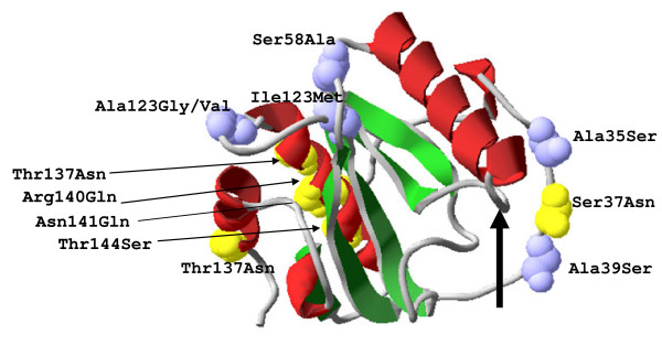
Figure 4.
Structural model of PGRP-S2 and PGRP-S3 proteins. Three-dimensional (3D) structural localization of mutated amino acids represented as yellow and blue (Van de Walls spheres). The PGRP domain has three α helices (red), five β strands (green) and coils (grey); Arrow indicates the specificity-determining residues responsible for the muramyl pentapeptide - MPP-Dap recognition.

