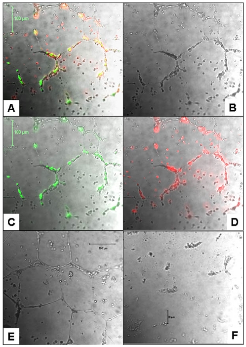
Figure 6. Functional analysis of long term expanded CB AC133+ cells to form tubes in Matrigel.
Tube like structures after 24 h of CB AC133+ cells and HDMVECs co-culture (A-D). Complete tubes in matrigel formed by HDMVECs incubated in the presence of CB AC133+ cells' supernatants (w/o EPCs and VEGF) for 24 h (E). When plated alone, CB AC133+ did not form tube like structures (F). Note in panels A-D, HDMVECs labeled with Calcein (C, green fluorescence) and CB AC133+ cells labeled with DiI (D, red fluorescence) co-localized (yellow; panel A) to form tube like structure. Most of the green fluorescent cells appeared to be structural part of the tubes, while some of the red florescence cells that did not became part of the tube network remained scattered between the tubes. Overlays of bright light microscopy and fluorescent microscopy images (A, C, D). Bright light microscopy only, images shown in panels B, E and F. Magnification 10x.

