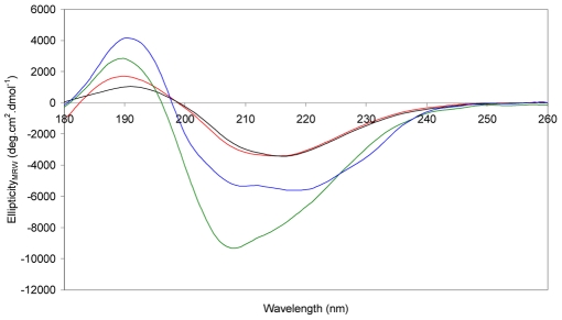
Figure 5. Circular dichroism spectra showing the structural transitions that occur in amyloid fibril formation.
Correctly folded MstnPP dimer (blue); MstnPP soluble aggregates before acidification (green); prefibrillar aggregates after overnight incubation in pH 5.3 at 60°C (red); mixture of prefibrillar aggregates and fibrils after one week incubation (black).

