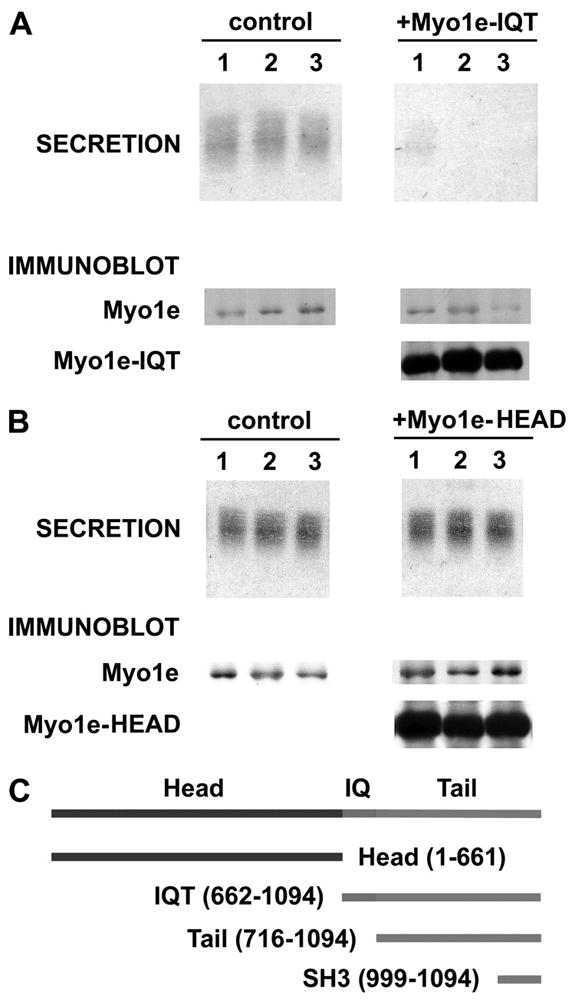
FIGURE 4. The IQT fragment of Myo1e inhibits cortical granule exocytosis, whereas the head domain is not inhibitory.

A, oocytes overexpressing the Myo1e-IQT show a nearly complete inhibition of cortical granule exocytosis. The secretion data show Coomassie-stained cortical granule lectin released from three separate control oocytes (in response to PMA) along with the inhibition of this release in three cells expressing Myo1e-IQT. The immunoblot data show that Myo1e is present in control oocytes and in cells expressing Myo1e-IQT. However, only the cells injected with Myo1e-IQT mRNA express this species (~45 kDa). B, oocytes that overexpress Myo1e-head show no reduction of cortical granule exocytosis. The secretion data show representative results from three controls and three oocytes overexpressing Myo1e-head (as documented by the immunoblot results, which are organized as in A). Overall, in n = 14 oocytes that exhibited a 12.9 ± 1.9-fold increase in the level of Myo1e-head relative to the native Myo1e, the secretion of cortical granule lectin was 106 ± 10% of control. C, schematic of Myole constructs used in this paper.
