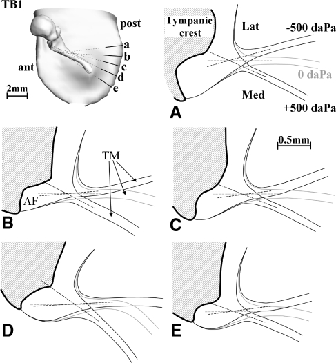
FIG. 4.
Cross-sections perpendicular to the annulus plane and orthogonal to the annulus boundary for −500, 0, and +500 daPa show the deformation at the peripheral boundary for TB1 (right ear). The location and orientation of the sections are indicated on the 3D models in the upper left corner. The dashed lines represent the midline of the TM at the edges just before it merges with the AF. When extrapolated, the lines for different pressures intersect at a single point. The point is situated at the base of the triangular thickening zone of the TM where it blends into the AF.

