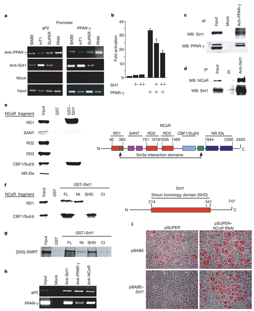
Figure 3.
Sirt1 reduces fat accumulation by repressing PPAR-γ activity through Sirt1–NCoR interactions. a, ChIP assays on 3T3-L1 cells infected as in Fig. 1c. Cells were differentiated for 7 days and assayed (Methods). b, Luciferase assays of 293T cells transfected with mouse PPAR-γ2 and pGL3–PPRE and with increasing concentrations of mouse Sirt1. Luciferase was measured 24 h later and was corrected for transfection efficiency. Data represent mean + s.e.m. of triplicate experiments carried out in duplicate. Asterisk indicates a statistical difference (P < 0.05). c, Co-immunoprecipitation of endogenous PPAR-γ and Sirt1 proteins in whole-cell extracts prepared from uninfected differentiated 3T3-L1 adipocytes. d, Co-immunoprecipitation of endogenous Sirt1 and NCoR proteins in HEK293 whole-cell extracts using pre-immune (PI) or anti-Sirt1 serum. e, Mapping the Sirt1 interaction interface of NCoR. Constructs encoding the indicated NCoR fragments were 35S-methionine-translated and used for GST pull-down experiments as previously described30. A schematic representation of NCoR functional domains is shown on the right. NR IDs, nuclear receptor interaction domains. SANT; Swi3, Ada2, NCoR, TF11B shared motif. f, Mapping the NCoR interaction interface of Sirt1. GST pull-down experiments were conducted using radiolabelled NCoR RD1 and CBF1/Su(H) interaction domains and the indicated fragments of Sirt1 fused to GST. A schematic representation of full-length Sirt1 is shown on the right. FL, full length; Nt, N-terminal; Ct, C-terminal. g, GST pull-down between the indicated fragments of Sirt1 fused to GST and 35S-methionine-translated SMRT. h, ChIP assay in uninfected 3T3-L1 cells differentiated for 7 days with insulin, IBMX and dexamethasone. i, RNAi of NCoR. 3T3-L1 cells were infected with either pBABE or pBABE–Sirt1 and co-infected with either pSUPER or pSUPER–NCoR RNAi. Puromycin-selected cells were then differentiated with insulin, IBMX and dexamethasone and stained with Oil red O 7 days later.

