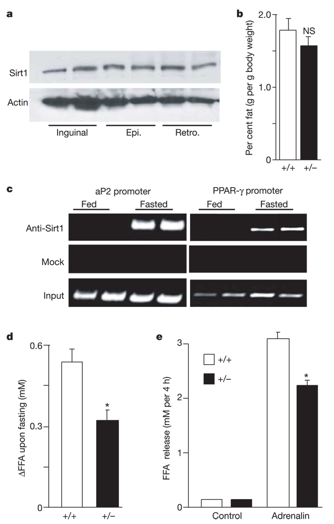
Figure 4.
Sirt1 promotes fat mobilization in vivo. a, Protein levels of Sirt1 in subcutaneous inguinal and visceral epididymal (Epi.) and retroperitoneal (Retro.) WAT depots in wild-type fed mice. Actin levels are shown as loading controls. Each lane represents an individual mouse. b, Weight of epididymal WAT depots in fed Sirt1 +/+ and Sirt1 +/− mice. Bars represent mean + s.e.m. (n = 10–12; NS, not significant). c, In vivo ChiP assay in FVB mice either fed or fasted overnight. Each lane represents an individual mouse. Findings represent five independent experiments done in duplicate. d, Change in FFA levels in blood from age-matched male Sirt1 +/+ and Sirt1 +/− mice between before and after overnight fasting. Bars represent mean + s.e.m. of 27–30 animals. Asterisk indicates a statistical difference from wild-type littermates (P < 0.05). e, Isolated adipocytes from Sirt1 +/+ and Sirt1 +/− mice incubated with or without adrenalin (10−5 M). After 4 h of incubation, FFA released in the medium were measured. Bars represent mean + s.e.m. of triplicate experiments. Asterisk indicates a statistical difference from Sirt1 +/+ adipocytes (P < 0.05).

