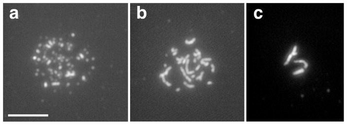
Figure 2.
Linear elements containing Rec10 protein. Spreads of meiotic nuclei were stained using an antibody to Rec10 protein. Dots (a), filaments (b), and bundles (c) appear to occur in that order during meiosis. The filaments and bundles appear to reflect the linear elements seen by electron microscopy (see text). Bar indicates 5 μm. Figure supplied by J. Loidl.

