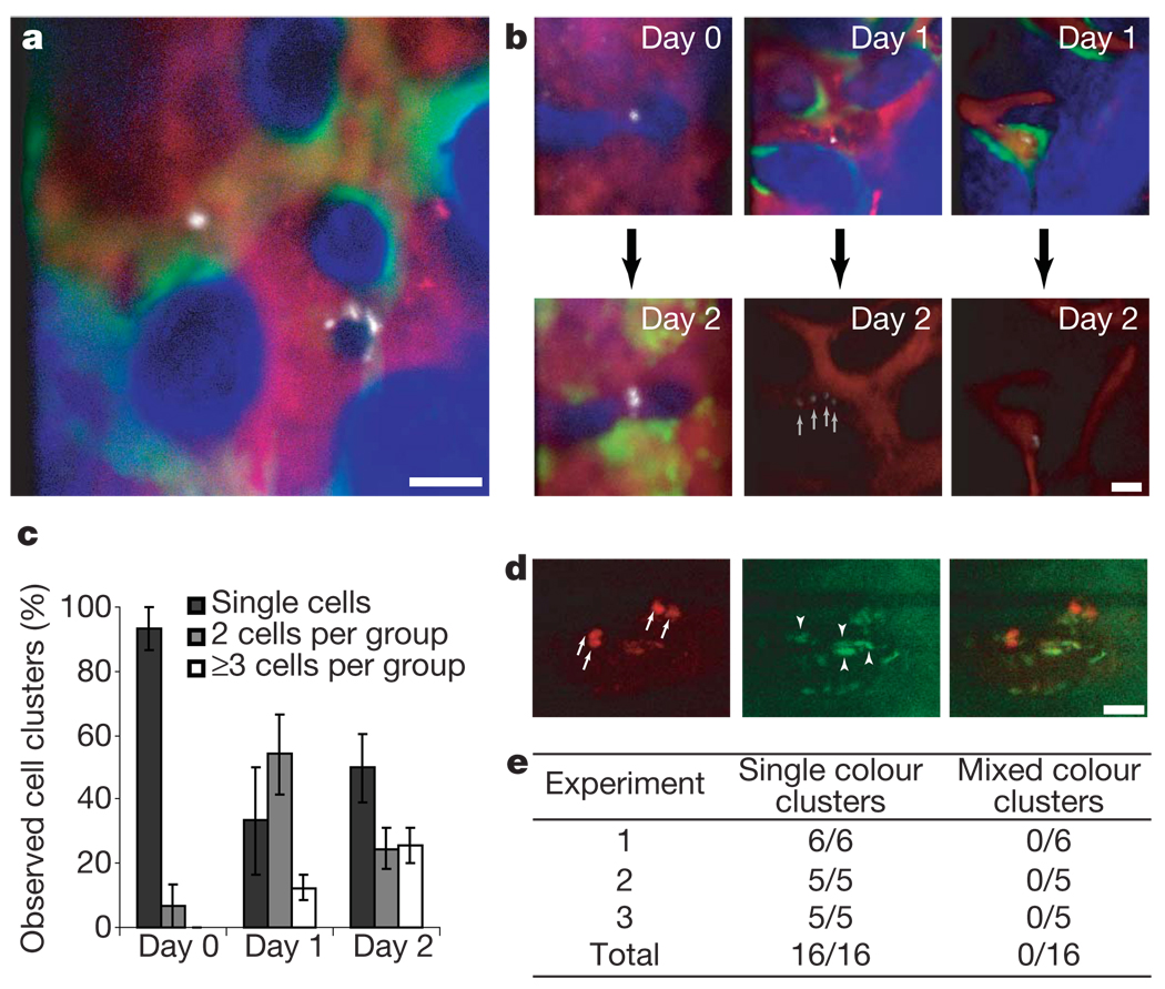
Figure 3. Engraftment is initiated by asynchronous HSPC cell divisions.
a, HSPC progeny were imaged 1 day after injection in irradiated recipients (n = 4 mice), revealing heterogeneity in cell clustering. Blue, bone; red, vasculature; green, osteoblasts; white, HSPC progeny. b, Cells were tracked from day 0 to day 2 (n = 2 recipient mice) or from day 1 to day 2 (n = 3 recipient mice) and diverse kinetics of cell division were observed. c, Increasing numbers of clusters containing 2 or ≥3 cells were observed in the days after injection (n = 4; error bars indicate s.e.m.). d, When 50% of cells were stained with DiD and 50% with DiI before injection, only single-colour clusters were observed. Red, DiD; green, autofluorescence. Arrows point at each DiD-positive cell within two clusters; arrowheads point at autofluorescent cells. Cells accepted for assessment had a dye/autofluorescence signal ratio >2 (in this example 2.82, 3.71, 4.12, 8.22). The same analysis was used to validate DiI signals. Scale bars in a, b, d are 50 µm. e, Summary of observed cell clusters in three independent experiments.

