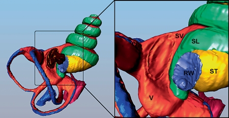
Fig. 3.
Reconstructed 3D anatomy of the fluid spaces of the guinea pig inner ear derived by segmentation of an OPFOS (orthogonal-plane fluorescence optical sectioning) image set [84] using Amira software. The enlargement shows the basal turn with the stapes removed (leaving an imprint of the footplate). V = Vestibule; SL = spiral ligament; RW = round window. The SL follows the periphery of the RW almost half way around, providing a major route for drugs in the ST near the RW to diffuse across into the vestibule. This anatomic pathway accounts for how drugs applied intratympanically to the RW can gain access to vestibular structures. Blue = Endolymph; orange = perilymph of SV and V; yellow = perilymph of ST; green = spiral ligament; red = sensory structures; purple = RW; magenta = cochlear aqueduct; brown = stapes.

