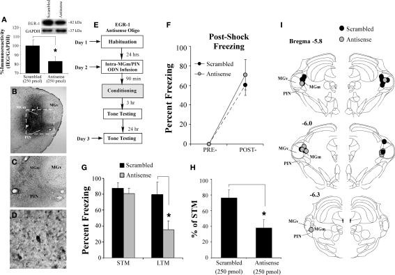
Figure 5.
Intra-MGm/PIN infusion of an EGR-1 antisense ODN impairs fear memory consolidation. (A) Western blot analysis of EGR-1 protein in the MGm/PIN of rats given intra-MGm/PIN infusion of scrambled ODN (n = 10) or EGR-1 antisense ODN (n = 10). Representative blots can be seen in the inset. *p < 0.05 relative to the scrambled ODN group. (B) Representative 4X photomicrograph of an animal given intra-MGm/PIN infusion of a biotinylated EGR-1 ODN (0.5 μl; 250 pmol) and sacrificed 30 min later. Note that the ODN diffusion is largely restricted to the MGm and PIN, and spares the MGv. (C–D) Higher level (20 and 40X, respectively) magnifications of MGm/PIN neurons containing biotinylated EGR-1 ODN label from the box in (B). Note the large number of cells exhibiting uptake of the ODN. (E) Schematic of the behavioral protocol. (F) Post-shock freezing scores in rats infused with either scrambled ODN (250 pmol, n = 9) or EGR-1 antisense ODN (250 pmol, n = 5) immediately after the conditioning trial. (G) Auditory fear memory assessed at both 3 and 24 h after fear conditioning in each group. The black bars represent the scrambled ODN-infused groups, while the gray bars represent the EGR-1 antisense ODN-infused groups. *p < 0.05 relative to the scrambled ODN-infused groups. (H) Data depicting LTM as a percentage of STM for each rat in each group. *p < 0.05 relative to the scrambled ODN-infused groups. (I) Cannula placements for rats infused with scrambled ODN (black circles) or EGR-1 antisense ODN (gray circles). Panels adapted from Paxinos and Watson (1997).

