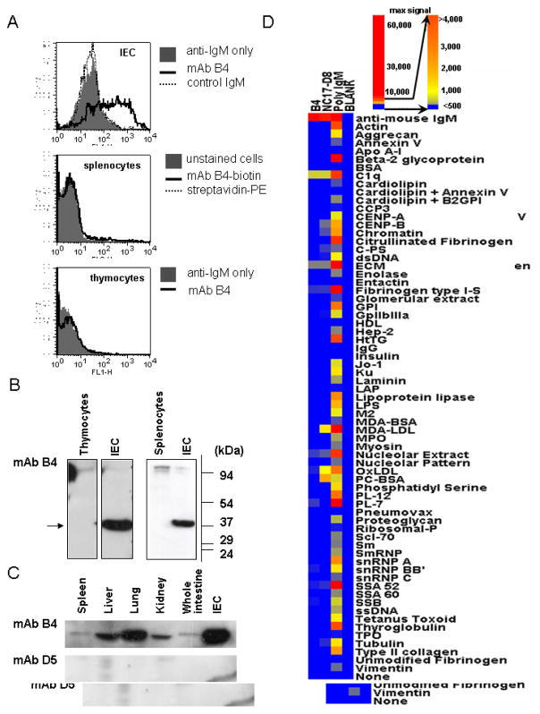
Figure 1.
Characterization of mAb B4 epitope expression. A, Representative flow cytometric histograms show binding of mAb B4 to a single cell suspension of IEC (top), splenocytes (middle) and thymocytes (bottom). Cells were incubated with mAb B4, and bound Ab was detected by using anti-mouse IgM (μ-chain specific) for IEC and thymocytes. In the case of splenocytes, mAb B4 labeled with biotin was used. B and C, Monoclonal Ab B4 epitope expression was determined by Western blot analysis of isolated from thymus, spleen or intestine cell in (B) or lysates were prepared from whole organs in (C). Data are representative of two independent experiments. D, Binding of mAb B4 to proteins typically found as targets of polyreactive natural Abs was studied by micro-array analysis. The positive control for mAb B4 binding are C1q and anti-mouse IgM antibody, positive controls for the micro-array are mAb NC-17D8 and polyclonal IgM, and the negative control is designated blank.

