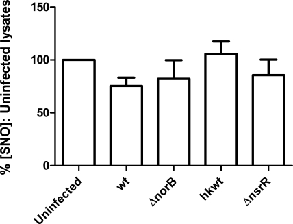
Figure 5.
Effect of inhibiting iNOS activity at the point of infection. J774.2 cells were activated with 1 μg/ml LPS and 1000 U/ml rmIFN-γ for 18 h, then infected with a suspension of log-phase N. meningitidis in fresh medium containing 100 μM 1400W. Strains were identical to those in Fig. 4. Lysates were produced using SNO-compatible lysis buffer plus 2% saponin. SNO content was determined by duplicate injection into I3− reaction mixture linked to ozone-based chemiluminescence and normalized to the protein concentration of each lysate. Data are percentages of SNO concentration in uninfected lysates. Bars denote means + se.

