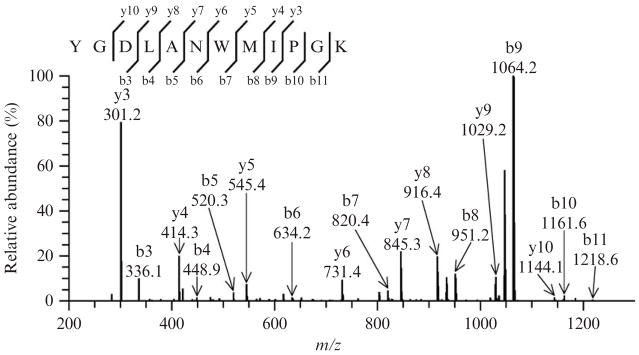
Figure 15.1.
Tandem mass spectrum of the untreated synthetic peptide YGDLANW-MIPGK ([M+2H]2+ ion at m/z 683.4) displaying a distinctive fragmentation pattern, characterized by the dominance of two ions, y3 and b9. Such a profile was interpreted to be due to the “proline effect,” which favors fragmentation on its amino terminal bond and suppresses other ions. For all spectra, ion annotation is based on results presented in the Sequest’s “display ion view” window, corroborated by the dta file.

