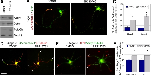
Figure 7.
Inhibition of GSK3β results in increased levels of tubulin PTMs and disrupts the polarized sorting of CA-Kinesin-1 and JIP1 in polarized stage 3 and unpolarized stage 2 cells. (A–C) Effects of GSK3β inhibition in polarized stage 3 cells. (A) Stage 3 cortical neurons were treated for 8 h with 10 μM SB216763. Cell lysates were analyzed by Western blotting with antibodies to acetylated α-tubulin, detyrosinated α-tubulin, polyglutamylated tubulin, or total β-tubulin. (B) Stage 3 hippocampal neurons were treated for 2–4 h with DMSO or 5–10 μM SB216763. The cells were then transfected with CA-Kinesin-mCherry and soluble YFP, allowed to express the exogenous proteins under additional treatment for 4–5 h, and then fixed and imaged. (C) Quantification of the percentage of stage 3 minor neurites or stage 5 dendrites that have accumulated CA-Kinesin-mCherry after treatment described in B. (D–F) Effects of GSK3β inhibition in unpolarized stage 2 cells. (D) Stage 2 hippocampal neurons expressing CA-Kinesin-1-mCit since the time of plating were treated for 8 h with DMSO or 10 μM SB216763 and then fixed and stained for total β-tubulin. (E) Stage 2 hippocampal neurons were treated for 8 h with DMSO or 10 μM SB216763 and then fixed and stained with antibodies to JIP1 and acetylated α-tubulin. (F) Quantification of the percentage of stage 2 neurites with CA-Kinesin-1 (D) or JIP1 (E) accumulated at the tips after treatment with DMSO or SB216763. Bars, 20 μm. Error bars, SEM. t test: *p < 0.01 compared with control DMSO-treated cells.

