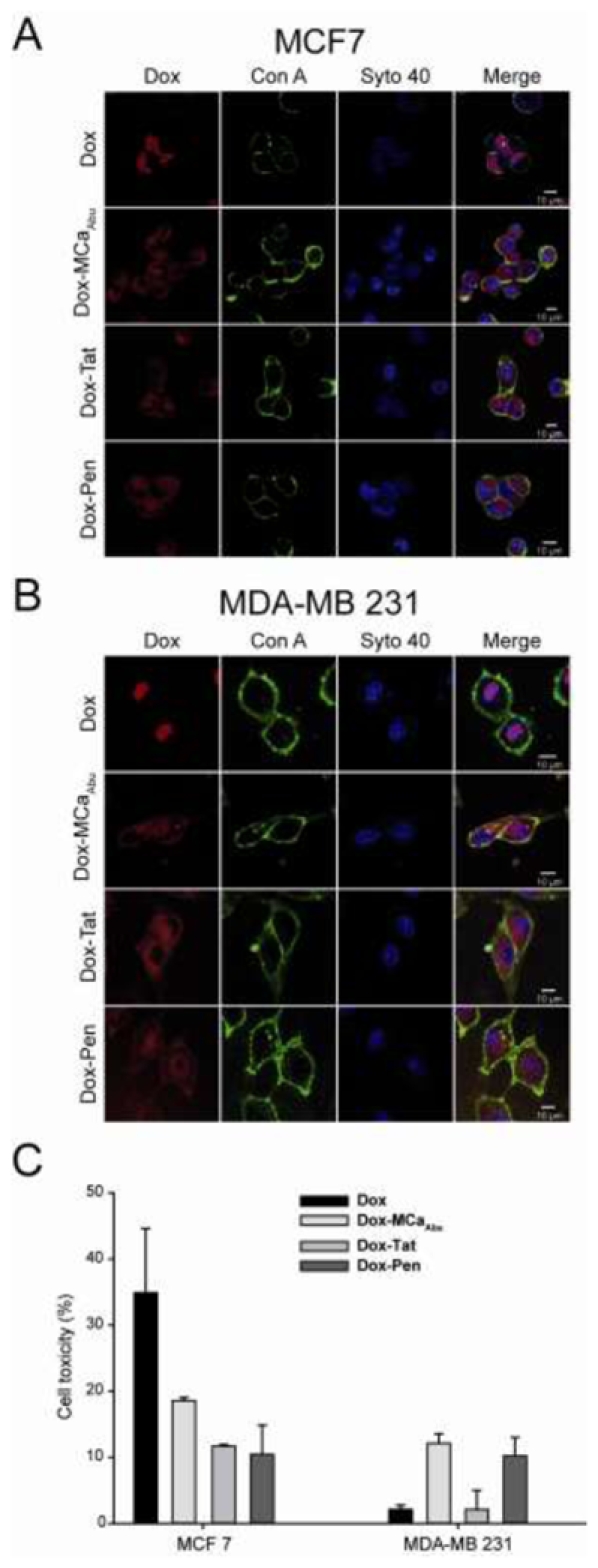
Figure 5.

Subcellular localization and cytotoxicity of free or conjugated Dox in MCF7 and MDA-MB 231 cells after short time treatments. (A) Confocal images of living MCF7 cells comparing the distribution of Dox fluorescence for free Dox (upper panels) or CPP-conjugated Dox (three lower panels) after a 2 hrs incubation. Cells were incubated with a drug concentration of 5 μM. (B) Same as in (A) but for MDA-MB 231 cells. (C) Cell toxicity of Dox and Dox-CPP conjugates evaluated with the MTT test in the same experimental conditions as in (A) and (B).
