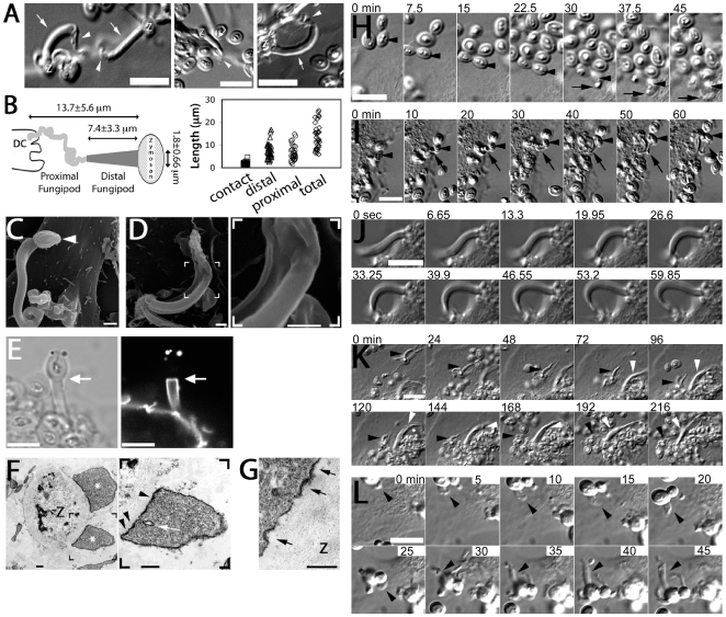
Figure 1. Morphology and formation of fungipods.
(A) DIC images of representative fungipods on the dorsal surfaces of immature DC after 4 hours exposure to zymosan. “Z” denotes the location of the fungipod attached zymosan particle. Arrows and arrowheads designate distal and proximal fungipod regions, respectively. Bars = 10 µm. (B) Schematic of fungipod morphology and distribution of measured contact site widths as well as lengths of distal, proximal and total fungipod regions (N = 35). (C) SEM image of a typical fungipod with roughly cylindrical distal geometry (9500x). Arrowhead denotes the attached zymosan particle. Bar = 1 µm. (D) SEM image of a fungipod with ribbon-like distal geometry displaying longitudinal ridges. Right panel shows a higher magnification view of the flattened structure in the distal fungipod from the bracketed area of the left panel. Left panel, 7500x; Right panel, 25000x, Bar = 1 µm. (E) DiI-labeled membrane of a distal fungipod at the zymosan contact site (arrow) viewed as a medial confocal section of the distal fungipod. (left, DIC; right, confocal fluorescence). Bars = 5 µm. (F) A fungipod/zymosan contact site seen in thin section TEM imaging. The right panel displays the left panel bracketed region at higher magnification. Designations: “Z”, Zymosan; “*”, distal fungipod; arrow, example of vesicle inside distal fungipod; arrowheads, examples of pits/membrane densities at the contact site. Left panel, 2700x; Right panel, 6500x; Bars = 500 nm. (G) Thin section TEM of membrane invaginations at a zymosan contact site displaying studded juxtamembrane densities. 11000x, Bar = 500 nm. (H) DIC imaging of the initial attachment and nascence of a fungipod. Arrowheads and arrows designate a zymosan particle associated with fungipod generation and the nascent fungipod, respectively. Times indicate elapsed time of DC attachment for the indicated zymosan particle. Bar = 10 µm. (I) DIC imaging of the maturation of a fungipod. Arrowheads and arrows designate a relevant zymosan particle and the growing fungipod, respectively. Times indicate elapsed time since the advent of the fungipod. Bar = 10 µm. (J) DIC imaging of the growth of a mature fungipod. Bar = 10 µm. (K) DIC imaging of fungipods (arrowheads) associated with zymosan and DC for several hours. Bar = 10 µm. (L) DIC imaging of a fungipod (arrowhead) formed by a DC in response to live S. cerevisiae. Bar = 10 µm.

